
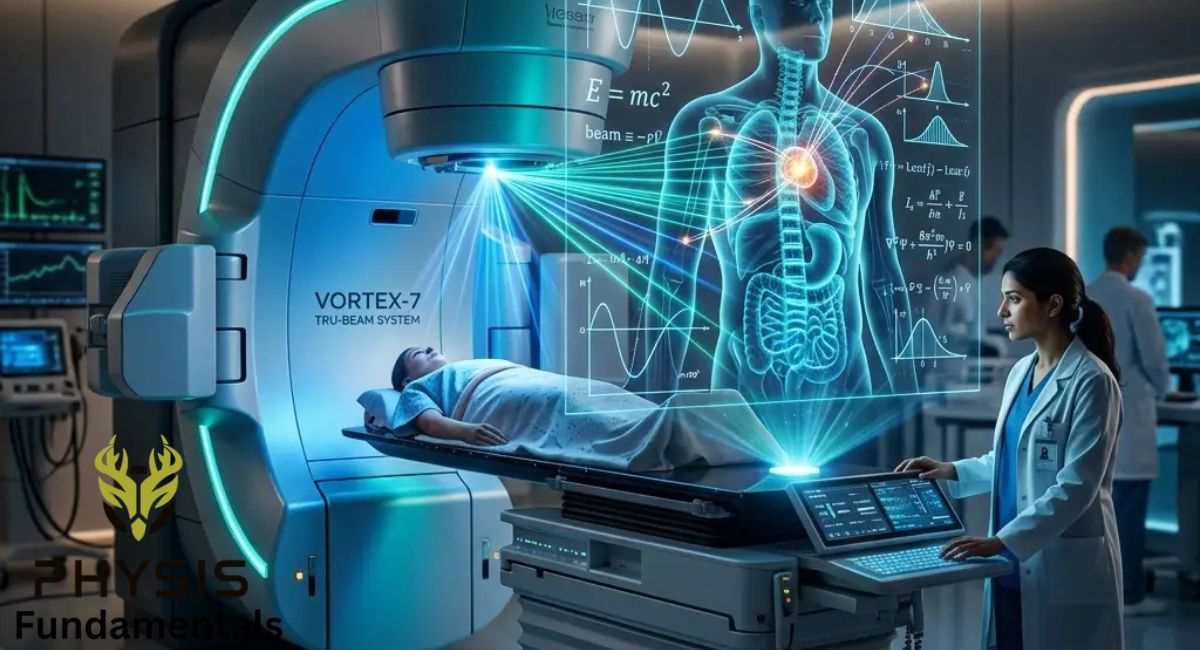
Medical physics is the science of applying physics principles to healthcare for the diagnosis, treatment, and prevention of diseases while ensuring the safe use of medical technology.
Medical physics that applies physical principles and techniques to medicine, especially in the diagnosis and treatment of diseases. It combines knowledge of physics with medical science for the development and use of technologies. This helps doctors to see inside the human body and treat patients safely and effectively. Medical physics plays an important role in modern healthcare technologies by improving the accuracy of medical imaging, reducing radiation risks, and enhancing treatment methods. In our daily life, medical physics is commonly used in hospitals and diagnostic centers through technologies such as X-ray imaging, ultrasound scans, CT-scans, and PET-scans. These techniques allow doctors to detect fractures, diagnose tumors, and to study internal organs without surgery. Thus, medical physics directly contributes to early diagnosis, effective treatment, and improved quality of life. We will study how positron emission tomography (PET) works through positron–electron annihilation and gamma-ray detection. The chapter will also explain the piezoelectric effect and its role in generating and detecting ultrasound waves for medical diagnosis. Furthermore, we will explore the production and use of X-rays.
Introduction to Radiations Used in Medical Physics.
Radiation is the transfer of energy through space or matter in the form of particles . In medical physics, radiation is mainly used for treatment. Radiations are broadly classified into non-ionizing diagnostics and ionizing radiations have enough energy to remove electrons from atoms, producing ions. Non-ionizing radiation does not have enough energy to remove electrons from atoms. waves or (imaging) and radiations. Examples of radiations used in medical physics are X-rays, gamma rays, and positrons. Interaction of X-rays and gamma rays with matter occurs through photoelectric effect and Compton scattering. The positrons are emitted from radioactive isotopes. On the other hand, examples of non-ionizing radiation are ultrasound waves and radio waves. These waves are reflected, refracted, or absorbed when interacting with the matter. Gamma rays, X-rays, ultrasonic waves, and radio waves are widely used in medical physics for diagnosis and treatment. X-rays internal organs, while commonly used to image bones and positrons, are used in nuclear medicine, such as gamma rays Single Photon Emission Computer Tomography, Positron Emission Tomography scans and cancer treatment, due to their high penetrating power. Ultrasonic waves are used in ultrasound imaging to examine soft tissues, monitor fetal diseases, and study internal organs safely without ionizing radiation. Radio waves are used in techniques such as MRI, where they help produce detailed images of internal body structures by interacting with atomic nuclei, making them especially useful for imaging soft tissues without harmful radiation exposure.
How Medical Physics Works.
Medical physics begins with understanding how energy interacts with the human body. Different forms of energy, such as X-rays, gamma rays, radio waves, magnetic fields, and sound waves, behave differently inside body tissues.
Medical physicists study these interactions to develop techniques that create detailed images of organs or precisely target diseased cells. They also calculate radiation doses, test medical machines, and ensure every procedure meets strict safety standards.
For example, during radiation therapy, a medical physicist carefully calculates how much radiation should be delivered to a tumor. The goal is to destroy cancer cells while protecting healthy tissues surrounding the tumor.
What Is Ultrasound?
Ultrasound is the use of high-frequency sound waves to produce images of organs, tissues, blood vessels, and developing babies inside the human body.
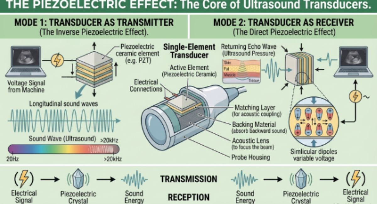
Ultrasound is a widely used non-invasive medical imaging technique that allows visualization of internal body structures such as tissues, organs, and the fetus during pregnancy. This imaging method operates on a physical principle known as the piezoelectric effect, which is central to the working of an ultrasound transducer. The transducer plays a dual role; it generates ultrasound waves and detects the reflected signals from inside the body. These sound waves have frequencies greater than 20,000 Hz (20 kHz), which is above the hearing range of the human ear.

How Does Ultrasound Work?
The ultrasound machine contains a device called a transducer. Inside the transducer are tiny piezoelectric crystals.
Step 1: Electrical Pulse
The ultrasound machine sends a short electrical pulse to the piezoelectric crystal.
↓
Step 2: Crystal Vibrates
Due to the inverse piezoelectric effect, the crystal rapidly expands and contracts.
↓
Step 3: Ultrasound Waves Produced
These vibrations generate high-frequency ultrasound waves.
↓
Step 4: Waves Enter the Body
The sound waves travel through skin, muscles, organs, and body fluids.
↓
Step 5: Reflection of Echoes
Whenever the waves encounter different tissues, part of the sound reflects back as echoes.
↓
Step 6: Crystal Receives Echoes
The returning echoes strike the same piezoelectric crystal.
↓
Step 7: Electrical Signals Produced
Using the direct piezoelectric effect, the crystal converts the echoes into electrical signals.
↓
Step 8: Image Formation
A computer processes these signals and displays a real-time image on the monitor.
The Piezoelectric Effect.
The piezoelectric effect is the property of certain crystals that convert mechanical energy into electrical energy and electrical energy into mechanical vibrations.
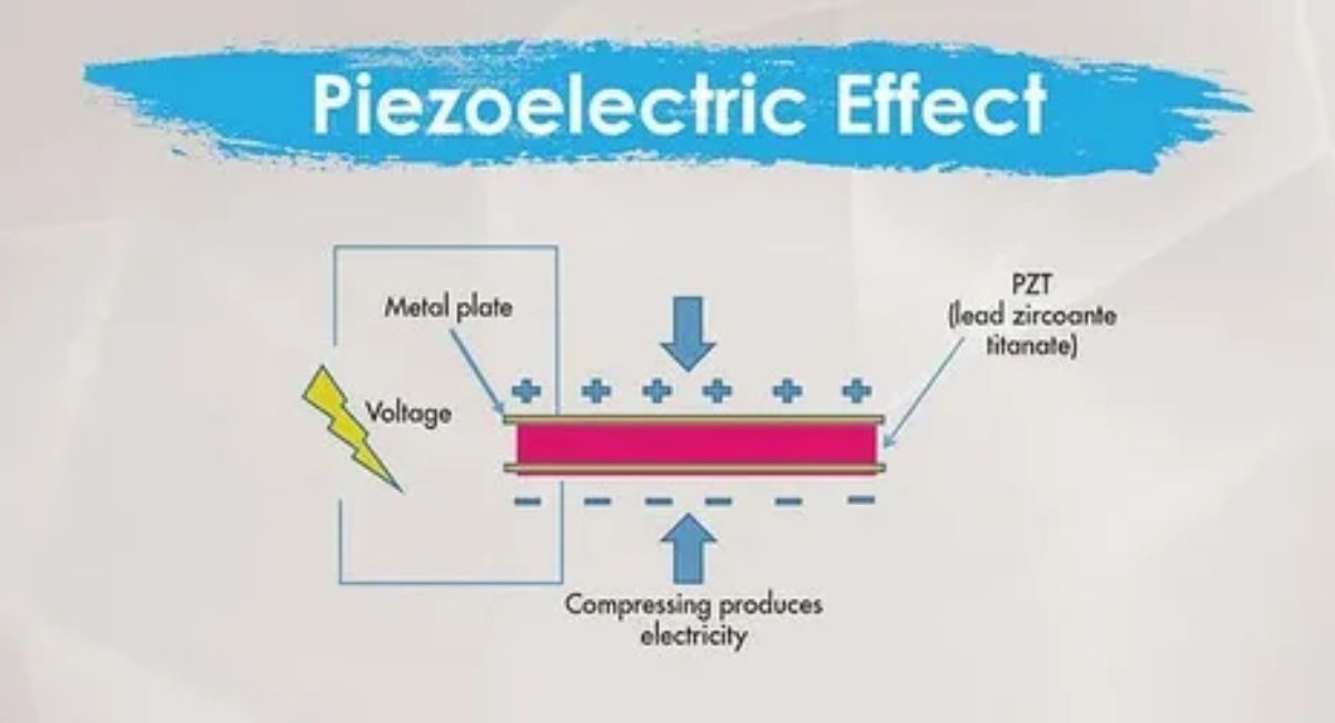
The word “piezein,” “piezoelectric” comes from the Greek word “to press” to the production of electricity. This effect was discovered in Curie 1880 . They were experimenting with crystals such as , and “electric” refers The piezoelectric effect makes it possible to turn pressure into voltage and vice versa. This is how ultrasound “talks” to the body. by two French brothers, Pierre Curie quartz, tourmaline and Jacques , and salt (sodium potassium tartrate). They found that when mechanical pressure was applied to these crystals, they generated an electric charge. Their experiments proved a strong connection between Rochelle mechanical force and electrical response The piezoelectric effect is possible because of the in certain crystals. unique internal structure crystals. There are two types of piezoelectric effect; direct piezoelectric effect and these crystals have no Centre of symmetry, and inside them, there are small regions with positive and negative charges. Normally, these charges are balanced, but when pressure is applied, the balance is disturbed, and the charges move slightly, causing an electric field to appear on the surface. This is called the direct piezoelectric effect. On the other hand, if we apply an electric field to the crystal, it changes shape a little. This is called the converse piezoelectric effect. A transducer generates ultrasound waves by the converse effect (an applied alternate voltage makes the crystal vibrate) and detects the returning echoes, the direct effect.
Piezoelectric Materials.
Common piezoelectric materials include:
- Quartz
- Lead Zircon ate Titanite (PZT)
- Barium Titanite
- Rochelle Salt
- Tourmaline
- Zinc Oxide
Among these, Lead Zirconate Titanite (PZT) is the most widely used material in modern ultrasound probes because it produces strong ultrasonic waves and is highly sensitive.
Types of Piezoelectric Effect.
Direct Piezoelectric Effect.
Mechanical pressure applied to a crystal generates an electrical voltage.
Example:
Pressing a quartz crystal produces a measurable electric signal.
- Pressure sensors
- Electronic weighing scales
- Microphones
- Medical ultrasound receivers
Inverse Piezoelectric Effect.
Applying an electric voltage causes the crystal to expand and contract rapidly, producing vibrations that generate ultrasonic sound waves.
- Ultrasound probes
- Sonar systems
- Medical imaging
- Ultrasonic cleaners
DIAGNOSTIC USE OF ULTRASOUND IN MEDICAL IMAGING.
Ultrasound is a widely used medical imaging technique that allows doctors to view internal body structures in real time. It operates by sending high frequency sound waves, typically in the range of 1 MHz-15 MHz, into the body using a device called a transducer. These waves travel through the body and reflect off boundaries between different tissues. The reflected echoes are then collected and analyzed to create an image. One of the key advantages of ultrasound is that it does not use ionizing radiation, making it a safe, noninvasive, and repeatable imaging method. The functioning of ultrasound imaging is based on several physical principles. First, the piezoelectric transducer emits pulses of ultrasound waves, which travel at different speeds depending on the type of tissue they pass through. When these waves encounter a boundary between two tissues with different acoustic impedances (determined by tissue density and sound speed), part of Screen Controls Cable connection Skin surface Transducer probe Skin Tissue Medical Physics the wave is reflected. The transducer then switches to receive mode and detects these reflected waves using the direct piezoelectric effect. These echoes are converted into electrical signals and processed by the machine. Finally, a computer calculates the time delay and intensity of the returning echoes to construct a visual image. Areas with stronger reflections appear brighter in the image, helping to reveal the internal structures of organs and tissues. Do you know? The crystals in an ultrasound probe are smaller than a grain of rice, but they can both send and detect sound waves.
Principle of Diagnostic Ultrasound
The working principle of ultrasound is based on the reflection of sound waves.
When ultrasound waves travel through the body, they pass easily through fluids and soft tissues. However, when they reach the boundary between different tissues—such as muscle and bone or fluid and soft tissue—part of the sound wave reflects back toward the transducer.
The ultrasound machine measures:
- The time taken for the echo to return.
- The strength of the reflected echo.
- The location from which the echo originated.
How Diagnostic Ultrasound Works.
The imaging process involves several simple steps.
-
Application of Ultrasound Gel
A clear, water-based gel is applied to the patient’s skin. The gel removes air between the skin and the transducer because air blocks ultrasound waves.
-
Transmission of Sound Waves
The transducer contains piezoelectric crystals. When electrical energy is applied, these crystals vibrate and produce high-frequency ultrasound waves.
-
Propagation Through the Body
The sound waves travel through muscles, organs, blood vessels, and body fluids.
-
Reflection of Echoes
Whenever the waves encounter different tissues, part of the sound energy is reflected back to the transducer.
-
Detection of Echoes
The same piezoelectric crystals receive the returning echoes and convert them into electrical signals.
-
Image Formation
A computer processes these electrical signals and displays real-time images on a monitor.
Types of Diagnostic Ultrasound
Different ultrasound techniques are designed for specific medical purposes.
Obstetric Ultrasound
This is one of the most familiar uses of ultrasound. It is used during pregnancy to monitor the developing baby.
Doctors use obstetric ultrasound to:
- Confirm pregnancy.
- Monitor fetal growth.
- Observe fetal movements.
- Detecting birth defects.
- Determine the baby’s position.
- Measure the amount of amniotic fluid.
Abdominal Ultrasound
Abdominal ultrasound examines organs such as:
- Liver
- Gallbladder
- Pancreas
- Kidneys
- Spleen
- Urinary bladder
It helps diagnose:
- Gallstones
- Kidney stones
- Liver disease
- Tumors
- Cysts
- Internal infections
Echocardiography (Heart Ultrasound)
Echocardiography uses ultrasound to examine the heart.
It helps doctors evaluate:
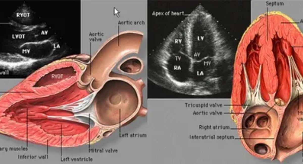
- Heart chambers
- Heart valves
- Pumping efficiency
- Blood circulation
- Congenital heart defects
Doppler Ultrasound
Doppler ultrasound measures the movement of blood through blood vessels.
It is used to diagnose:
- Blood clots
- Narrowed arteries
- Blocked blood vessels
- Poor circulation
- Heart valve disorders
-
Musculoskeletal Ultrasound
This technique examines:
- Muscles
- Tendons
- Ligaments
- Joints
- Soft tissues
It is commonly used for sports injuries and tendon tears.
-
Thyroid Ultrasound
Doctors use thyroid ultrasound to detect:
-
- Thyroid nodules
- Thyroid enlargement
- Thyroid cysts
- Tumors
- Inflammation
-
Breast Ultrasound
Breast ultrasound helps distinguish between fluid-filled cysts and solid breast masses. It is often used alongside mammography for a more complete evaluation.
Diagnostic Applications of Ultrasound.
Ultrasound plays a vital role in diagnosing many medical conditions.
Pregnancy Monitoring
Ultrasound allows doctors to monitor fetal growth, heartbeat, and development while ensuring the health of both mother and baby.
Heart Disease
Echocardiography helps diagnose heart valve diseases, heart failure, congenital defects, and abnormal heart function.
Liver Disorders
Ultrasound can identify fatty liver disease, tumors, cysts, liver enlargement, and cirrhosis.
Kidney Disorders
It detects kidney stones, tumors, cysts, urinary obstruction, and infections.
Gallbladder Diseases
Ultrasound is considered the first-choice imaging technique for detecting gallstones and gallbladder inflammation.
Blood Vessel Disorders
Doppler ultrasound evaluates blood circulation and identifies narrowed arteries, aneurysms, and blood clots.
Soft Tissue Injuries
Muscles, tendons, ligaments, and joints can be examined quickly without surgery.
Advantages of Diagnostic Ultrasound
Diagnostic ultrasound offers many benefits in healthcare.
- Completely non-invasive.
- Painless and comfortable.
- Does not use ionizing radiation.
- Safe for pregnant women and unborn babies.
- Produces real-time images.
- Portable and easy to use.
- Less expensive than CT or MRI.
- Can be repeated frequently without significant health risks.
X-RAYS.
X-rays are high-energy electromagnetic waves that pass through the body to create images of bones, organs, and other internal structures.
Discovery of X-Rays.
X-rays were discovered in 1895 by the German physicist Wilhelm Conrad Rontgen while experimenting with cathode rays.
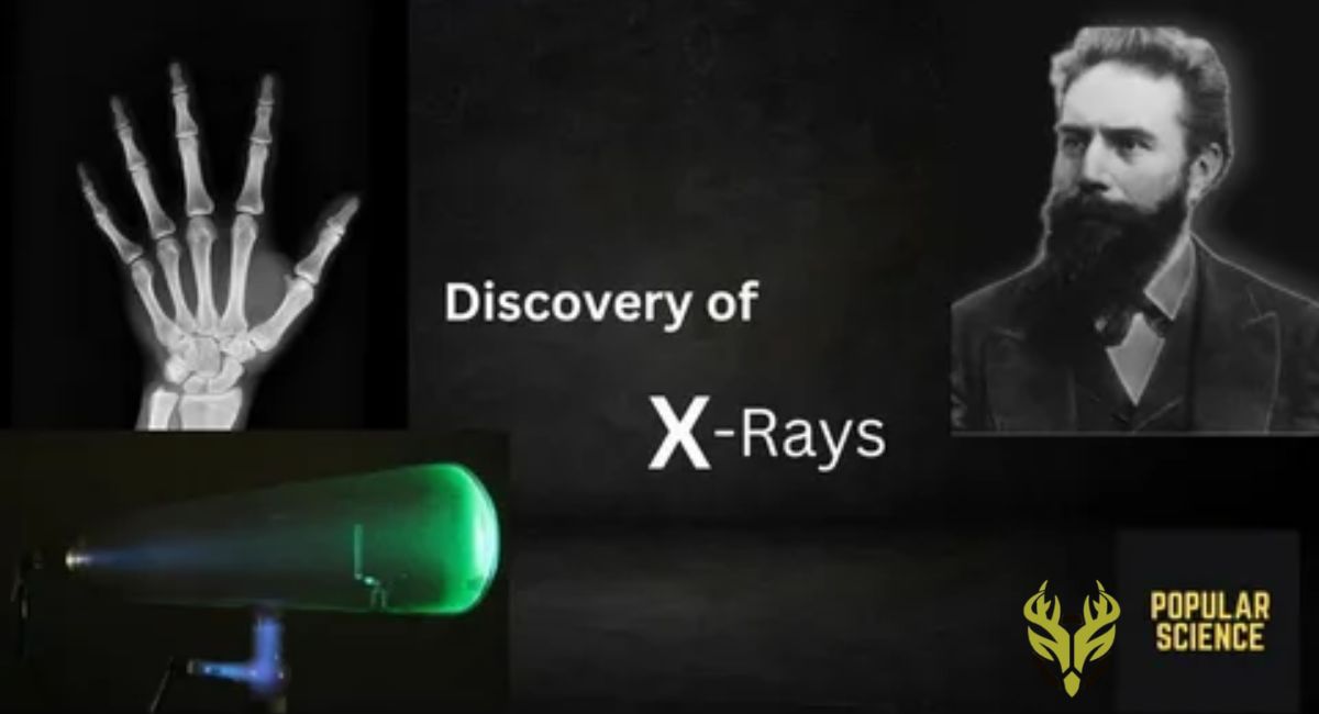
During his experiment, he noticed that an unknown type of radiation could pass through objects and produce images on a fluorescent screen. Since the nature of the radiation was unknown, he called them X-rays, where “X” means unknown. Soon afterward, Rontgen produced the world’s first medical X-ray image—an image of his wife’s hand showing her bones and wedding ring. This discovery transformed medicine and earned him the first Nobel Prize in Physics in 1901.
Properties of X-Rays.
X-rays have several unique characteristics.
- They are invisible to the human eye.
- They travel at the speed of light.
- They have very short wavelengths.
- They possess high energy and high penetrating power.
- They can pass through soft tissues but are absorbed more by dense materials like bones.
- They can ionize atoms and molecules.
- They can expose photographic film and digital detectors.
- They travel in straight lines.
- They are not affected by electric or magnetic fields.
How Are X-Rays Produced?
X-rays are generated inside a machine called an X-ray tube.
The production process involves several steps:
Step 1: Heating the Cathode
An electric current heats a metal filament inside the cathode, causing it to release electrons.
Step 2: Acceleration of Electrons
A high voltage accelerates the electrons toward the positively charged metal target (anode).
Step 3: Collision with the Target
The fast-moving electrons strike the tungsten target at very high speeds.
Step 4: Production of X-Rays
The sudden deceleration of electrons produces high-energy X-rays.
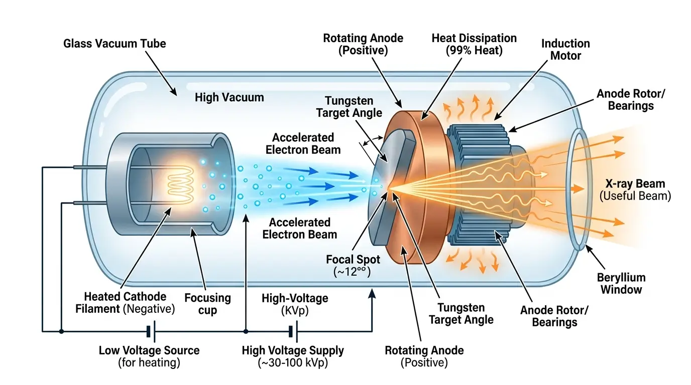
Only about 1% of the electrical energy is converted into X-rays, while the remaining 99% becomes heat.
How Do X-Rays Form an Image?
When X-rays pass through the body, different tissues absorb different amounts of radiation.
- Bones absorb most X-rays and appear white.
- Muscles and organs absorb some X-rays and appear gray.
- Air-filled spaces, such as the lungs, absorb very little radiation and appear black.
A digital detector or photographic film records the transmitted X-rays, creating an image that doctors can interpret.
Types of X-Rays.
-
Diagnostic X-Rays
These X-rays are used to diagnose diseases and injuries.
-
- Chest X-rays
- Dental X-rays
- Bone X-rays
- Mammography
- Fluoroscopy
-
Therapeutic X-Rays
High-energy X-rays are used in radiation therapy to destroy cancer cells while minimizing damage to nearby healthy tissues.
Medical Uses of X-Rays
X-rays are essential in many areas of healthcare.
Diagnosing Bone Fractures
Doctors use X-rays to identify broken or cracked bones after accidents or falls.
Dental Imaging
Dentists use X-rays to detect cavities, tooth infections, impacted teeth, and jaw problems.
Chest Examination
Chest X-rays help diagnose:
- Pneumonia
- Tuberculosis
- Lung cancer
- Heart enlargement
- Fluid around the lungs
Mammography
Special X-rays are used to detect breast cancer at an early stage.
Detecting Arthritis
X-rays reveal changes in bones and joints caused by arthritis.
Monitoring Medical Devices.
Doctors use X-rays to check the position of pacemakers, artificial joints, screws, and implants after surgery.
Non-Medical Uses of X-Rays
X-rays are valuable outside the medical field as well.
Airport Security
Security scanners inspect luggage for prohibited items.
Industrial Inspection
Engineers use X-rays to detect hidden cracks or defects in metal structures and machinery.
Scientific Research
Scientists study the internal structure of crystals and materials using X-ray diffraction.
Art Restoration
Experts examine paintings and historical artifacts without damaging them.
Advantages of X-Rays
X-rays provide many benefits.
- Fast and painless examination.
- Non-invasive diagnostic technique.
- Excellent for detecting bone injuries.
- Widely available in hospitals and clinics.
- Relatively inexpensive compared with CT and MRI.
- Helps detect diseases at an early stage.
- Guides doctors during certain medical procedures.
How Are Characteristic X-Rays Produced?
To understand how characteristic X-rays are produced, we first need to look at the arrangement of electrons inside an atom.
In hydrogen and other light elements, electrons occupy energy levels that are relatively close together. When an electron moves from a higher energy level to a lower one, it releases a small amount of energy as electromagnetic radiation. Depending on the energy difference, the emitted light falls in the infrared, visible, or ultraviolet region of the electromagnetic spectrum.
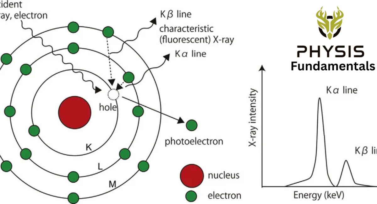
However, the situation is different in heavy elements such as tungsten, which is commonly used in X-ray tubes. In these atoms, electrons are arranged in several concentric shells around the nucleus, known as the K, L, M, N, and O shells. The K-shell is the innermost shell and lies closest to the nucleus, followed by the L-shell, M-shell, and so on.
The electrons in the inner shells are held very tightly by the strong positive charge of the nucleus. Therefore, a large amount of energy is required to remove an electron from one of these inner shells.
When a high-energy electron strikes a heavy atom, it can knock out an electron from the K-shell, leaving an empty space called a vacancy. This makes the atom unstable. To regain stability, an electron from a higher-energy shell, such as the L-shell or M-shell, moves down to fill the vacancy in the K-shell.
As the electron drops to the lower energy level, it releases the difference in energy as a high-energy photon. Because the energy difference between the inner shells of heavy atoms is very large, the emitted photon has enough energy to be an X-ray.
Since each element has its own unique arrangement of electron energy levels, the emitted X-rays have specific wavelengths and frequencies that are unique to that element. For this reason, these X-rays are called characteristic X-rays because they are characteristic of the atom from which they originate.
Step-by-Step Process of Characteristic X-Ray Production
- A high-speed electron strikes a heavy atom (such as tungsten).
- An inner-shell electron, usually from the K-shell, is ejected.
- The atom becomes unstable because of the vacancy in the inner shell.
- An electron from a higher shell (L, M, or N) moves down to fill the vacancy.
- The energy difference between the two shells is released as a high-energy photon.
- This photon is emitted as a characteristic X-ray.
Characteristic X-Ray Equations
When an inner-shell electron is removed, an electron from a higher shell falls into the vacancy. The energy released is emitted as an X-ray photon.
The energy of the emitted X-ray is given by:
E=hν=hc/λ
- E = Energy of the X-ray photon
- h = Planck’s constant (6.626×10⁻³⁴ J)
- ν = Frequency of the X-ray
- c = Speed of light (3.0×10^8 m/s)
- λ = Wavelength of the X-ray
Kα (K-alpha) X-Rays
A Kα X-ray is produced when an electron from the L-shell (n = 2) falls into a vacancy in the K-shell (n = 1).
Transition
L→K
Energy Equation
EKα=EK−EL
where:
- EK = Energy of the K-shell
- EL = Energy of the L-shell
Since the K-shell is much lower in energy than the L-shell, the energy difference is emitted as a Kα X-ray photon.
Kβ (K-beta) X-Rays
A Kβ X-ray is produced when an electron from the M-shell (n = 3) falls into the K-shell (n = 1).
Transition
M→K
Energy Equation
EKβ=EK−EM
where:
- EK = Energy of the K-shell
- EM = Energy of the M-shell
Because the M-shell is farther from the nucleus than the L-shell, the energy difference is larger. Therefore:
EKβ>EKα
This means Kβ X-rays have higher energy and a shorter wavelength than Kα X-rays.
Lα (L-alpha) X-Rays
An Lα X-ray is produced when an electron from the M-shell falls into a vacancy in the L-shell.
Transition
M→L
Energy Equation
ELα=EL−EM
Lβ (L-beta) X-Rays
An Lβ X-ray is produced when an electron from the N-shell falls into a vacancy in the L-shell.
Transition
N→L
Energy Equation
ELβ=EL−EN
Summary of Characteristic X-Ray Transitions
| X-Ray Series | Electron Transition | Energy Equation |
| Kα | L → K | EKα=EK−EL |
| Kβ | M → K | EKβ=EK−EM |
| Lα | M → L | ELα=EL−EM |
| Lβ | N → L | ELβ=EL−EN |
Production of X-rays
It consists of a high vacuum tube called X-ray tube. When the cathode C is heated by the filament F, it emits electrons which are accelerated towards the anode T. If V is the potential difference between C and T, the (K.E.) with which the electron strikes the target is given by max ( K.E. ) = Ve max Suppose that these fast moving electrons of energy Ve strike a target made of tungsten or any other heavy element. It is possible that in a collision, the electrons in the innermost shells, such as K or L, will be knocked out. Suppose that one of the electrons in the K- shell is removed, thereby producing a vacancy or hole in that shell. The electron from the L-shell jumps to occupy the hole in the K-shell, thereby emitting a photon of energy.
h , called the K X-ray given by a
hfKα = EL- E K
It is also possible that the electron from the M-shell might also jump to occupy the hole in the K-shell. The photons emitted are K X-ray with energies of β. These photons give rise to K X-ray and so on. Β
hfKβ = EM– EK
Intensity =hc/eV limn 0 0.04 K The photons emitted in such transitions i.e., inner shell transitions are called characteristic X-rays, because their energies depend upon the type of target material. The holes created in the L and M-shells are occupied by transitions of electrons from higher energy states creating more X-rays. The characteristic X-rays appear as discrete lines on a continuous spectrum.
What Is the Continuous X-Ray Spectrum?
The continuous X-ray spectrum is produced when fast-moving electrons are rapidly slowed down by the metal target, causing them to release energy in the form of X-ray photons. This process is called Bremsstrahlung or braking radiation.
How Is Bremsstrahlung Radiation Produced?
The production of Bremsstrahlung X-rays occurs in several steps.
Step 1: Electrons Are Accelerated
Inside the X-ray tube, a high voltage accelerates electrons toward the metal target at very high speeds.
Step 2: Electrons Strike the Target
The fast-moving electrons collide with the atoms of the target material.
Step 3: Sudden Deceleration
As the electrons pass close to the positively charged nuclei of the target atoms, they are strongly attracted and rapidly slow down or change direction.
Step 4: X-Ray Emission
Whenever a charged particle slows down or accelerates, it emits electromagnetic radiation. In this case, the rapidly decelerating electrons release their lost kinetic energy as X-ray photons.
This emitted radiation is called Bremsstrahlung (braking radiation).
Why Is It Called a Continuous Spectrum?
Not every electron loses the same amount of energy.
- Some electrons lose all of their kinetic energy in a single collision, producing a high-energy X-ray with the shortest possible wavelength.
- Other electrons lose only part of their energy during each collision. They continue moving and undergo several collisions before stopping completely.
- Each collision produces an X-ray photon with a different energy.
As a result, X-rays with many different wavelengths and energies are produced, creating a continuous spectrum instead of a few discrete lines.
Maximum Energy of Bremsstrahlung X-Rays
If an electron loses all of its kinetic energy in a single collision, the entire kinetic energy is converted into one X-ray photon.
The maximum photon energy is given by:
Emax=hfmax
where:
Emax = Maximum energy of the X-ray photon
- h = Planck’s constant
- f max = Maximum frequency of the emitted X-ray
Since the electron’s kinetic energy is
KE=eV
the maximum X-ray energy becomes
eV=hfmax
where:
- e = Charge of an electron
- V = Accelerating voltage
Minimum Wavelength
The shortest wavelength produced is called the minimum wavelength λmin).
Using
E=hc/λ
we obtain
λmin=hc/eV
where:
- h = Planck’s constant
- c = Speed of light
- e = Electron charge
- V = Accelerating voltage
This equation shows that increasing the accelerating voltage produces X-rays with higher energy and shorter wavelengths.
Characteristics of the Continuous X-Ray Spectrum
- Produced by the sudden slowing down of fast-moving electrons.
- Also called Bremsstrahlung or braking radiation.
- Contains X-rays with a wide range of wavelengths.
- Forms a continuous spectrum rather than separate spectral lines.
- Higher tube voltage produces shorter wavelength X-rays.
- Accounts for most of the X-rays generated in a diagnostic X-ray tube.
Real-Life Example
Imagine driving a car at high speed and suddenly applying the brakes. The car’s kinetic energy is rapidly lost as heat and sound. In a similar way, fast-moving electrons inside an X-ray tube are suddenly “braked” when they approach the nuclei of tungsten atoms. Instead of producing heat and sound alone, part of their lost kinetic energy is emitted as Bremsstrahlung X-rays. Because each electron slows down differently, the emitted X-rays have different energies, creating a continuous spectrum.
What Is X-Ray Imaging?
X-ray imaging is a diagnostic method that uses X-rays to create pictures of bones, organs, and other internal structures without surgery.
Ionization by X-Rays
X-rays carry enough energy to remove electrons from atoms. This process is called ionization. When an X-ray strikes an atom, it can knock out one of its electrons. The atom then becomes electrically charged, or ionized. Ionization transfers energy from the X-ray to the body’s tissues. This is why X-rays are called ionizing radiation, and why doctors use the lowest possible radiation dose needed to obtain a clear image.
Reduction in X-Ray Intensity.
As X-rays travel deeper into the body, fewer X-rays remain because some are absorbed or scattered by tissues. Therefore, the intensity of the X-ray beam decreases continuously as it passes through the body. The decrease follows an exponential pattern, meaning the beam loses a certain fraction of its intensity as it passes through each layer of tissue rather than the same fixed amount.
Image Intensifiers
In some medical procedures, doctors need to observe moving organs in real time. For example, they may watch blood flowing through blood vessels or guide a catheter during surgery. To make these live X-ray images brighter and clearer, special devices called image intensifiers are used. An image intensifier collects the weak X-ray image and converts it into a much brighter image that can be displayed on a monitor.
Benefits of Image Intensifiers
- Produce brighter images.
- Improve image quality.
- Allow doctors to see internal organs in real time.
- Reduce the amount of radiation needed for imaging.
- Improve patient safety.
Contrast in X-Ray Images.
Contrast refers to the difference in brightness between different tissues in an X-ray image.
Good contrast allows doctors to easily distinguish one structure from another.
For example:
- Bones appear white because they absorb many X-rays.
- Muscles and organs appear gray.
- Air inside the lungs appears black because it absorbs very few X-rays.
The better the contrast, the easier it is for doctors to identify injuries or diseases.
Hard X-Rays and Soft X-Rays.
The energy of X-rays affects how they are used in medicine.
Hard X-Rays
Hard X-rays have high energy and can penetrate dense materials such as bones.
They are mainly used for:
- Bone fractures
- Joint examinations
- Chest imaging
Soft X-Rays.
Soft X-rays have lower energy and are absorbed more easily by soft tissues.
They are commonly used for:
- Mammography (breast imaging)
- Examining soft tissues
- Detecting small abnormalities
Contrast Media.
Sometimes, certain organs or blood vessels are difficult to see on a normal X-ray because they absorb X-rays almost as much as the surrounding tissues.
To improve visibility, doctors use contrast media (also called contrast agents).
These are special substances that absorb X-rays more strongly than body tissues, making the targeted area appear more clearly on the image.
The two most common contrast media are:
- Iodine-based contrast agents
- Barium sulfate
Depending on the examination, the contrast medium may be:
- Injected into a vein.
- Swallowed as a liquid.
- Introduced into a body cavity.
Common Uses of Contrast Media
- Examining blood vessels (angiography)
- Imaging the digestive system
- Detecting kidney and urinary tract disorders
- Studying the stomach and intestines
- Identifying blockages or tumors
COMPUTED TOMOGRAPHY SCANNING (CT-SCAN)
A Computed Tomography (CT) scan is a medical imaging technique that uses multiple X-ray images taken from different angles and combines them with computer technology to create detailed cross-sectional and 3D images of the body’s internal organs, bones, blood vessels, and soft tissues.
Historical Background.
The CT scanner was developed in the early 1970s by Sir Godfrey Hounsfield of England and Allan Cormack of South Africa. Their groundbreaking invention transformed medical imaging by allowing doctors to view detailed cross-sectional images of the human body. In recognition of this achievement, they were awarded the 1979 Nobel Prize in Physiology or Medicine.
Why Is a CT Scan Needed?
A normal X-ray produces only a flat (2D) image. In this image, different structures such as bones, muscles, and organs overlap, making it difficult to examine them individually. A CT scan solves this problem by taking many X-ray images from different angles and combining them into thin slices. These slices provide a much clearer view of the body’s internal structures.
How Does a CT Scan Work?
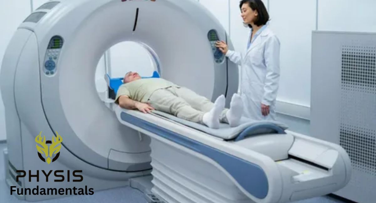
A CT scanner consists of an X-ray tube, detectors, a patient table, and a computer.
The scanning process occurs in several steps.
Step 1: The Patient Lies on the Table
The patient lies comfortably on a motorized table that slowly moves through the CT scanner.
Step 2: X-Ray Tube Rotates
The X-ray tube rotates around the patient’s body while emitting X-rays from many different angles.
Step 3: Detectors Receive the X-Rays
Special detectors on the opposite side measure the X-rays that pass through the body.
Step 4: Multiple Images Are Collected
Hundreds of X-ray images are captured from different directions in just a few seconds.
Step 5: Computer Creates Cross-Sectional Images
A powerful computer processes all the collected data and produces detailed cross-sectional slices of the body.
Step 6: Formation of a 3D Image
The computer combines all the slices to create a highly detailed three-dimensional (3D) image of the internal organs, bones, and tissues.
Why Is It Called Computed Tomography?
The name Computed Tomography (CT) comes from two words:
- Computed – A computer processes the X-ray data and converts it into images.
- Tomography – Derived from the Greek word “tomes,” meaning “slice.” It refers to producing thin cross-sectional images of the body.
Earlier, this technique was often called Computed Axial Tomography (CAT scan) because the images were taken along the body’s axis. Today, the term CT scan is more commonly used.
Main Components of a CT Scanner.
A CT scanner contains several important parts.
X-Ray Tube
Produces X-rays and rotates around the patient during scanning.
Detectors
Measure the X-rays that pass through different parts of the body.
Motorized Table
Moves the patient slowly through the scanner to capture images of different body sections.
Computer
Processes the X-ray data and reconstructs detailed cross-sectional and 3D images.
Applications of CT Scans
CT scans are used to diagnose and monitor many medical conditions.
Brain Imaging
CT scans help detect:
- Stroke
- Brain tumors
- Internal bleeding
- Head injuries
- Skull fractures
Chest Examination
Doctors use CT scans to diagnose:
- Lung cancer
- Pneumonia
- Blood clots
- Chest injuries
Abdominal Imaging
CT scans examine:
- Liver
- Kidneys
- Pancreas
- Spleen
- Gallbladder
- Intestines
Bone and Joint Injuries
CT scans detect:
- Complex fractures
- Joint injuries
- Bone tumors
Cancer Diagnosis
CT scans help doctors:
- Detect tumors
- Determine tumor size
- Monitor treatment progress
- Plan radiation therapy
Blood Vessel Imaging
With contrast media, CT scans can clearly show arteries and veins to detect aneurysms, blockages, or narrowed blood vessels.
Advantages of CT Scans
CT scans offer many advantages over traditional X-rays.
- Produce detailed cross-sectional images.
- Create three-dimensional (3D) images.
- Show bones, organs, blood vessels, and soft tissues clearly.
- Detect diseases at an early stage.
- Quickly identify internal injuries after accidents.
- Help doctors accurately locate tumors.
- Guide biopsies and minimally invasive procedures.
- Faster than MRI in many emergency situations.
Limitations of CT Scans
Although CT scans are highly useful, they also have some limitations.
- Use ionizing X-rays.
- Radiation dose is higher than a standard X-ray.
- Frequent CT scans should be avoided unless medically necessary.
- Contrast dye may cause allergic reactions in some patients.
- Not always recommended during pregnancy unless the benefits outweigh the risks.
Radiation Exposure
CT scans use X-rays, which are a form of ionizing radiation. Although the radiation dose is generally considered safe for medical use, it is higher than that of a standard X-ray.
Doctors carefully weigh the benefits against the small radiation risk before recommending a CT scan. Special care is taken when scanning children and pregnant women, as they are more sensitive to radiation.
CT Scan vs Ordinary X-Ray
| Feature | CT Scan | Ordinary X-Ray |
| Images | Cross-sectional and 3D | Flat 2D image |
| Detail | Very high | Moderate |
| Soft Tissues | Clearly visible | Less detailed |
| Bones | Excellent detail | Good detail |
| Scan Angles | Multiple directions | Single direction |
| Radiation Dose | Higher | Lower |
| Best Used For | Tumors, internal organs, trauma, brain injuries | Bone fractures, chest imaging |
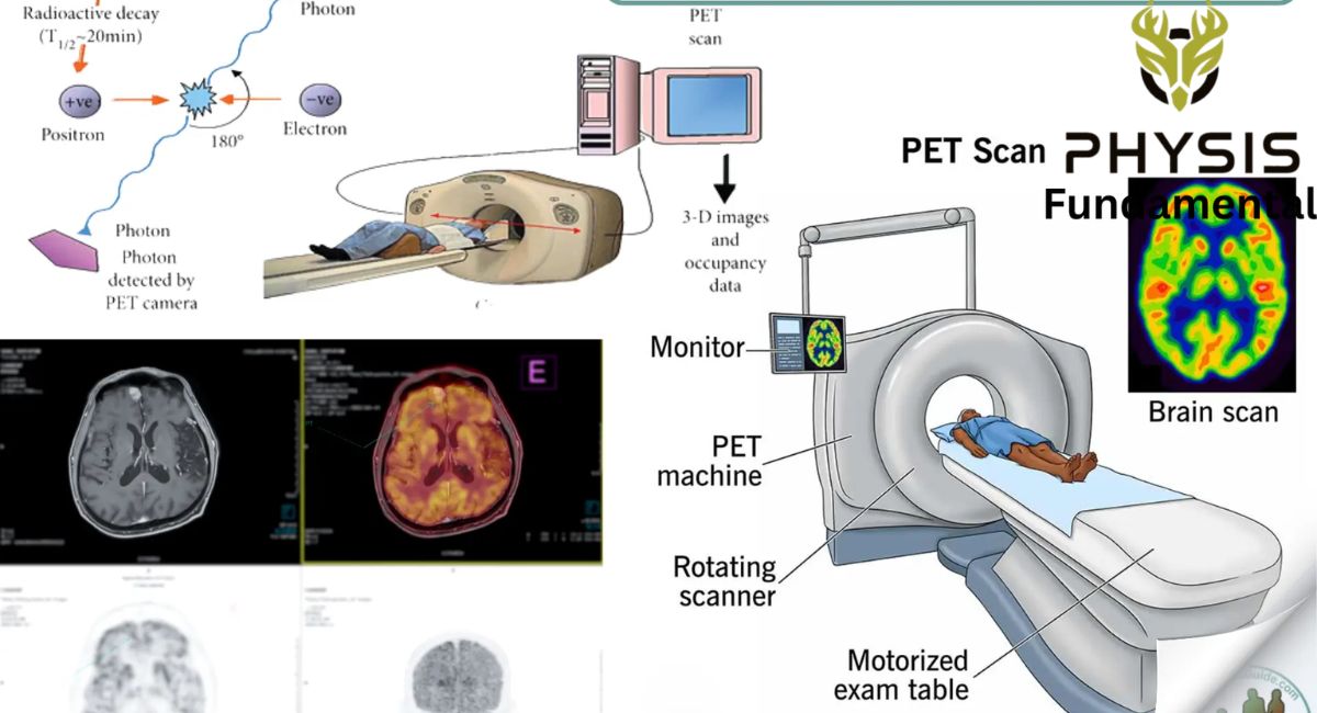
Positron Emission Tomography (PET) Scan
A PET scan is a non-invasive imaging technique that uses a small amount of radioactive tracer to show how organs and tissues are working inside the body.
Principle of a PET Scan
A PET scan works on the principle of positron emission and annihilation.
The radioactive tracer contains unstable atoms that release positrons (positively charged particles). When a positron meets an electron inside the body, both particles disappear in a process called annihilation.
During annihilation, their mass is converted into energy according to Einstein’s famous equation:
E=mc2
This energy is released as two gamma-ray photons that travel in opposite directions.
Special detectors surrounding the patient detect these gamma rays and send the information to a computer, which reconstructs detailed images of the tracer distribution inside the body.
How Does a PET Scan Work?
The PET scanning process involves several simple steps.
Step 1: Injection of the Radioactive Tracer
A very small amount of radioactive tracer is injected into a vein.
The most commonly used tracer is Fluorine-18 Fluorodeoxy glucose (FDG), which behaves like glucose (sugar).
Step 2: Tracer Travels Through the Body
The tracer circulates through the bloodstream and is absorbed by different organs and tissues.
Highly active cells, such as cancer cells, brain cells, and heart muscle cells, absorb more tracer because they require more energy.
Step 3: Positron Emission
The radioactive atoms inside the tracer are unstable.
As they decay, they emit positrons.
Step 4: Annihilation
Each positron travels only a very short distance before colliding with a nearby electron.
When they meet, both particles are destroyed in a process called annihilation.
Their mass is converted into energy, producing two gamma-ray photons that move in opposite directions.
Step 5: Detection of Gamma Rays
A ring of detectors surrounding the patient detects the two gamma rays at the same time.
This simultaneous detection allows the scanner to determine where the annihilation occurred.
Step 6: Image Formation
A powerful computer analyzes millions of these detected events and reconstructs detailed color images.
Bright areas on the PET image represent regions where more tracer has accumulated, indicating higher metabolic activity.
Components of a PET Scanner
A PET scanner consists of several important parts.
Radioactive Tracer
Produces positrons inside the body.
Detector Ring
Detects pairs of gamma rays emitted during annihilation.
Computer
Processes the detected signals and reconstructs detailed images.
Patient Table
Moves the patient smoothly through the scanner.
Why Do Cancer Cells Appear Bright?
Cancer cells grow and divide much faster than normal cells.
Because of their rapid growth, they consume more glucose for energy.
Since the PET tracer behaves like glucose, cancer cells absorb larger amounts of the tracer.
As a result, these areas emit more radiation and appear as bright spots on PET images.
Medical Applications of PET Scans
PET scans are widely used in many areas of medicine.
Cancer Diagnosis
PET scans help doctors:
- Detect cancer at an early stage.
- Determine the size and location of tumors.
- Identify whether cancer has spread to other organs.
- Monitor the effectiveness of cancer treatment.
- Detect the recurrence of cancer after treatment.
Brain Imaging
PET scans are used to study brain function and diagnose conditions such as:
- Alzheimer’s disease
- Parkinson’s disease
- Epilepsy
- Dementia
- Brain tumors
Heart Imaging
PET scans evaluate blood flow and heart muscle function.
Doctors use them to:
- Detects coronary artery disease.
- Identify damaged heart muscle after a heart attack.
- Assess heart function before surgery.
Medical Research
PET scans help researchers understand how organs function and how diseases develop, leading to improved treatments and new medicines.
Advantages of PET Scans.
PET scans provide several important benefits.
- Detect diseases before structural changes become visible.
- Show organ function rather than just anatomy.
- Identify cancer at an early stage.
- Monitor the success of treatment.
- Produce highly detailed functional images.
- Help doctors make more accurate diagnoses.
Limitations of PET Scans
Although PET scans are extremely useful, they have some limitations.
- They involve exposure to a small amount of ionizing radiation.
- They are more expensive than many other imaging techniques.
- Patients must receive a radioactive tracer before the scan.
- The equipment is available mainly in specialized hospitals.
- PET scans often need to be combined with CT or MRI for precise anatomical details.
PET Scan vs CT Scan
| Feature | PET Scan | CT Scan |
| Main Purpose | Shows organ function and metabolism | Shows body structure and anatomy |
| Radiation Source | Radioactive tracer inside the body | External X-rays |
| Images | Functional images | Structural images |
| Best Used For | Cancer, brain function, heart activity | Bones, organs, injuries, tumors |
| Early Disease Detection | Excellent | Moderate |
- What does a time difference in detecting gamma rays at two detectors indicate in a PET scan?
- Why is ultrasound not effective for imaging air-filled organs like the lungs? Explain.
- What happens when the X-ray contrast between two tissues is too low? Suggest a solution.
- How does Computed Tomography scanning reduce the overlapping of organs seen in plain X-rays? Explain.
- What could be a possible medical cause of uneven tracer concentration seen in a PET brain scan?
- What is the purpose of lead collimators in PET gamma-ray detectors?
- What is the piezoelectric effect, and how does it allow transducers to both emit and detect ultrasound waves?
- Describe how an ultrasound image is created using the concepts of echoes and reflected sound waves.
- Explain how attenuation of X-rays by different tissues creates contrast in the resulting image. Include an example.
- Compare CT and conventional X-ray imaging with respect to image clarity and diagnostic value.
- Write a brief note on PET. How is it used to scan malignant tissues? 20.6 How would you explain the inner shell transitions of X-rays? Also explain characteristic and continuous X-rays.
-
FAQs
- What is medical physics?
- Why is medical physics important in healthcare?
- What are the main applications of medical physics?
- What is the difference between ionizing and non-ionizing radiation?
- Which types of radiation are used in medical diagnosis?
- How do medical physicists ensure patient safety?
- What is ultrasound imaging?
- How does an ultrasound machine work?
- What is a transducer in ultrasound?
- Why is ultrasound considered a safe imaging technique?
- What is the frequency range of ultrasound used in medical imaging?
- Why is ultrasound gel applied before scanning?
- Why can’t ultrasound effectively image air-filled organs like the lungs?
- What are the main advantages of diagnostic ultrasound?
- What are the different types of ultrasound scans?
- What medical conditions can ultrasound diagnose?
- What is the piezoelectric effect?
- Who discovered the piezoelectric effect?
- What is the difference between the direct and inverse piezoelectric effects?
- How does the piezoelectric effect help produce ultrasound waves?
- Which materials are commonly used as piezoelectric crystals?
- Why is Lead Zirconate Titanite (PZT) widely used in ultrasound probes?
- What are the everyday applications of the piezoelectric effect?
- What are X-rays?
- Who discovered X-rays?
- How are X-rays produced?
- Why are X-rays considered ionizing radiation?
- How do X-rays create images of the human body?
- Why do bones appear white on an X-ray?
- What is the difference between hard X-rays and soft X-rays?
- What are characteristic X-rays?
- What is Bremsstrahlung (continuous) X-ray radiation?
- What are the medical applications of X-rays?
- What are the advantages and limitations of X-ray imaging?
- What are contrast media, and why are they used in X-ray imaging?
- What is a CT scan, and how does it work?
- What is the difference between a CT scan and a conventional X-ray?
- What are the advantages and disadvantages of CT scanning?
- What is a PET scan?
- How does a PET scan work?
- What is positron-electron annihilation?
- Why do cancer cells appear brighter on PET scans?
- What radioactive tracer is commonly used in PET scanning?
- What are the medical applications of PET scans?
- What is the difference between a PET scan and a CT scan?
- What are the advantages and limitations of PET imaging?
- Conclusion.
Medical physics sits at the intersection of physics and healthcare, turning fundamental scientific principles into life-saving diagnostic and therapeutic tools. As this overview of medical physics shows, each imaging technology relies on a distinct physical phenomenon: ultrasound depends on the piezoelectric effect to convert electrical energy into sound waves and back again; X-rays are produced through the deceleration of high-speed electrons, generating both characteristic and continuous (Bremsstrahlung) radiation; CT scanning combines multiple X-ray angles into detailed cross-sectional images; and PET scanning harnesses positron-electron annihilation to reveal metabolic activity at the cellular level.
Together, these applications of medical physics allow doctors to look inside the human body without invasive surgery — detecting fractures, tumors, heart conditions, and organ dysfunction earlier and more accurately than ever before. Understanding the physics behind these tools doesn’t just deepen scientific knowledge; it highlights how precise, evidence-based innovation directly improves patient outcomes, safety, and quality of life. As imaging technology continues to advance, medical physics will remain essential to the future of accurate diagnosis and effective treatment.